
Patella cubiti
A Bone That Might Not Exist
Six years of research unraveling a diagnosis that had concealed several distinct conditions for over a century
23.8.2026, MUDr. Vojtěch Kunc, Ph.D.
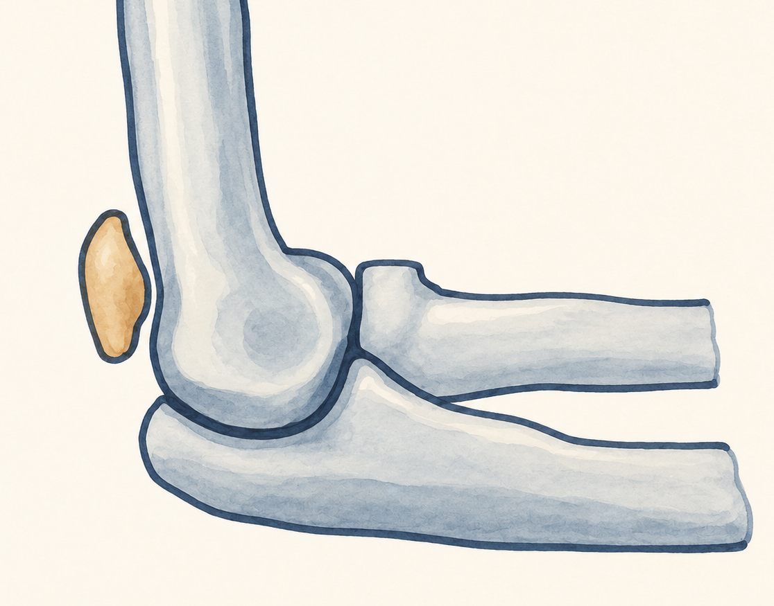
A separate bony structure may be visible on an X-ray at the back of the elbow. Is it a true accessory bone – a sort of “kneecap of the elbow"? A sesamoid bone within the triceps tendon? A congenital nonunion of the olecranon? Or the result of a previous injury?
Since 2020, we have been working to unravel this anatomical enigma. After several setbacks, we gradually came to realize that the answer was not a simple one, but rather a more precise map of rare conditions that had been hidden under a single term for generations.
Main findings: Of the 38 published cases described as patella cubiti , only three met our criteria. Even in these cases, however, the historical documentation does not allow for a definitive conclusion.
A catchy term that was never clearly defined
Patella cubiti literally means “kneecap of the elbow.” The concept is intriguing: an anatomical variant consisting of a separate bone at the back of the elbow joint, resembling the kneecap in both its position and function. The problem is that the term has been used in the literature to describe almost any separate bony structure near the olecranon seen on an X-ray whose origin was not immediately apparent.
As early as 1903, Kienböck’s description raised the question of whether this was a congenital anatomical variant or the result of an injury. Over the following decades, case reports continued to accumulate, but a consistent definition never emerged. Some patients had been aware of the bony structure since childhood and remained asymptomatic, while others presented after an injury with pain and loss of extension. In some cases, the bone was located within the triceps tendon; in others, it formed part of the articular surface, while still others appeared to represent old, ununited fracture fragments. Thus, the same term came to describe conditions that differed in both their anatomy and underlying cause.
Where did the search begin?
Our investigation into this condition began in 2020, while studying accessory bones of the elbow. At that time, we published a research about their prevalence, their location, and a modified classification. At the same time, we proposed three simple radiographic criteriawhich should be fulfilled by any accessory bone ovid shape, smooth margin and regular cortical to medullary bone ratio.
Patella cubiti was not included in the classification at the time. However, the question remained unresolved. The more historical radiographs and case descriptions we examined, the less it seemed that all the authors were describing the same condition. This was the point at which an anatomical curiosity turned into a detective story: we first had to look beyond the diagnosis reported in the original publication and reassess each case based on its anatomical features.
Thirty-eight cases from two centuries
We systematically searched scientific databases, reviewed the references of relevant publications, and examined articles retrieved from historical archives. Some case reports were nearly a century old. Many contained little more than a brief written description and a poor-quality reproduction of an old X-ray, often published in the authors’ native language.
The help of colleagues in interpreting texts written in different languages was essential to this research. Ultimately, we identified 31 studies describing 38 patients whose findings had originally been classified as patella cubiti. We then reassessed each case individually, examining the patient’s age, affected side, history of trauma, symptoms, bone morphology, anatomical relationship to the olecranon, elbow joint, and triceps tendon, as well as the treatment provided and its outcome.
Four main diagnoses
True patella cubiti
According to our definition, patella cubiti is a congenital intra-articular accessory bone that articulates with both the olecranon and the trochlea of the humerus, is covered with hyaline cartilage, and has no bony continuity with the rest of the ulna. Its presence should alter the geometry of trochlear notch of the ulna (incisura trochlearis) . It is precisely this relationship with the joint that distinguishes it from a bone located solely within the tendon.
Os sesamoideum tricipitale
This sesamoid bone is located extra-articularly within the distal tendon of the triceps brachii muscle. It may appear similar on a plain X-ray, but it does not contribute to articular congruence. From both an anatomical and biomechanical perspective, it represents an entirely distinct entity. Interestingly, it can fracture following trauma and cause significant symptoms.
Congenital olecranon pseudarthrosis
In this condition, there is no additional bone; instead, the olecranon is separated from the rest of the ulna by non-ossified tissue. Typical features include symptoms present since childhood, limited elbow extension, and the absence of a convincing history of trauma. Surgical reports from some historical cases described fibrocartilaginous tissue or a mobile pseudoarthrosis.
Traumatologic causes
Historically, the same term was also used to describe old fractures, avulsion injuries of the triceps tendon insertion, nonunions, persistent olecranon physes, and other accessory bones in the posterior elbow. When a fragment did not meet the criteria for a congenital accessory bone and there was a clear history of trauma, we considered a traumatic origin more likely.
The most important answer is an honest “we don’t know”
It would be tempting to conclude this story by stating that patella cubiti does not exist. However, our data do not support such a conclusion. Three historical cases are consistent with true patella cubiti . However, none of these cases provides sufficiently high-quality radiographs, MRI scans, or a complete anatomical description to establish the existence of this entity with certainty.
The most accurate conclusion is therefore somewhat different: even after more than a century, the literature has yet to conclusively establish the existence of true patella cubiti or definitively rule it out. But this does not mean that our investigation has failed. Quite the opposite. We have been able to distinguish several distinct conditions that had historically been grouped under a single diagnosis and establish a framework for evaluating future cases with much greater precision.
The heart of the enigma: The congenital, traumatic, and developmental theories are likely not three competing explanations for a single diagnosis. Rather, they describe several distinct conditions that have historically been grouped under the same name.
How should the diagnostic process look:
The first step is to obtain a high-quality, standardized lateral radiograph of the elbow. The separate bony structure should be assessed for three key features of an accessory bone: an oval shape, smooth margins, and a uniform corticomedullary ratio. The geometry of the trochlear notch of the ulna should also be evaluated, and the patient’s history of trauma carefully reviewed.
However, MRI should play a decisive role. It can reveal whether the bony structure is connected to the rest of the ulna, whether it is covered with articular cartilage, and whether it lies within the distal triceps tendon. These anatomical relationships are essential for distinguishing true intra-articular patella cubiti from a sesamoid bone, a pseudoarthrosis, or a post-traumatic fragment. Without this information, a plain radiograph will often remain just the beginning of the story.
Why is it clinically relevant?
This is not merely a matter of terminology. An asymptomatic congenital accessory bone, a sesamoid bone within the triceps tendon, a mobile pseudoarthrosis, and a post-traumatic nonunion are not interchangeable diagnoses. They differ in their anatomy, biomechanics, and likely treatment indications. Historical case reports describe both conservative and surgical management, but the cases are too heterogeneous to establish a reliable treatment algorithm.
The key clinical lesson is to exercise caution: not every bone posterior to the olecranon should be labelled patella cubiti, the underlying cause should not be inferred from a single radiograph, and a precise anatomical diagnosis should be established before deciding on treatment. In such rare conditions, accurate diagnosis may be the first meaningful step towards appropriate treatment.
Six years unraveling an anatomical mystery
What I appreciate most about this work is that it reflects a less glamorous, yet deeply rigorous side of scientific research. It is not about a large patient cohort or a novel implant. It is about patiently reviewing historical case reports, carefully examining every radiograph, tracking down original publications, and being willing to acknowledge that, in 19 cases, the available evidence simply does not allow us to reach a definitive conclusion.
Since 2020, a small bone at the edge of an X-ray has evolved into a six-year research project. Our aim was never to prove the existence of patella cubiti at all costs. Rather, it was to distinguish between rare conditions that had long been grouped under a single historical term, allowing their true differences to emerge. For me, this is the beauty of anatomical detective work: answers do not always come from spectacular findings, but from carefully distinguishing between things that merely appear similar.
This research is the result of a collaborative effort. The co-authors are Viktorie Adamcová, Matin Čepelík, Šárka Salavová and David Kachlík. This article would not have been possible without the contributions of colleagues who helped retrieve historical publications, translate texts, prepare illustrations, and revise the manuscript. When researching a topic spanning decades and multiple languages, such collaboration is just as important as the analysis itself.
Patella cubiti may indeed exist, but until we encounter a well-documented case with detailed anatomical findings and MRI evidence, it will remain one of the most fascinating unsolved mysteries of the elbow joint.
Publication:
Kunc V., Adamcová V., Čepelík M., Salavová Š., Kachlík D. Systematic review of patella cubiti: Does it really exist? Annals of Anatomy. 2026;267:152871. DOI 10.1016/j.aanat.2026.152871
Next to read: